
How does a CT scan work? A CT scan uses x-rays and computer software to produce a 2D or a 3D image of the section of the body being scanned.
CT stands for Computed Tomography. “Computed” means calculated, or worked out. “Tomography” is “imaging by sections using any kind of penetrating wave”. Tomography is made from two Greek words: “tomos”, which means slice or sections, and “grapho”, which means to write.
A CT scan is basically a system that takes multiple X-ray images of a section of the body and then uses computer software to put them together. They are different from an MRI in this respect because MRIs use a magnetic field to make the protons in the body align, which can then be used to produce an image. A CT scan rotates around, taking many X-ray images that can then be imaged as slices.
When you have a single x-ray image taken, the x-ray technician will have you stand or sit against an imaging plate. The x-ray “camera” is pointed at the part of you to be x-rayed and fired. A blast of X-rays are released and they fly into you. X-rays are fairly powerful and they can fly through many things, but they cannot travel through dense bone or dense things like tumors. They can fly through blood, skin, fat, muscle, and soft tissue without much trouble. The plate that the x-rays strike can then be used to make an image depending on the quantity of x-rays that have hit it. The image becomes a negative because it is dark where a lot of x-rays strike it and light or white where few strike it. Bones block the X-rays and appear white. Flesh doesn’t block many x-rays and it appears in shades of grey depending on its density.
A CT scan uses X-rays in the same way, but the machine takes many more of them and from many different angles. There are several different types of CT scanners, but the most common type has the device that produces the X-rays on a wheel that can rotate round the patient inside the scanner. On the opposite side is the image plate that is connected to a computer. Once the patient is inside, the machine is activated and it begins to rotate. The table the patient is lying in is motorized so they can be moved forwards and backwards without having to move the wheel itself. The beam of X-rays is very narrow, which gives the layers that make a CT scan so useful. As the wheel rotates round the patient, x-rays are fired from the X-ray tube and picked up by the image plate. Each time the X-rays are fired, the wheel has rotated on and the angle that they pass through the body changes. That means instead of just getting the one image of a bone or organ that you get with a regular X-ray, there are multiple angles of the same bone or organ. The X-rays hit the image plate and the computer is instantly able to log how many X-rays hit each part of the plate. This data can then be used to make an image of the body. There are so many angles that CT scanners are able to see hundreds of different densities. Most CT scanners can display this as a 2D image, but modern CT scanners can actually make it into a 3D image. The accuracy is so good that CT scans can be used to track internal bleeding or to find cancers.
Because they take so many images from so many different angles, they can be used to view organs and softer tissue that a regular X-ray would not be able to see. That makes them very useful. For example, it is possible to do a colonoscopy with a CT scan rather than having to insert a camera. However, because they take so many X-ray images, the patients do undergo a larger dose of radiation than with a regular X-ray. Depending on the type of CT scan, they can have between 100 and 1,000 times more radiation than a regular x-ray. If you have an x-ray of your abdomen, for example, you will be exposed to 0.7 millisieverts (mSv) and if you have a CT of your abdomen, you will be exposed to roughly 8 mSv. The x-ray radiation is ionizing and can therefore damage the cells in your body. CT scans are an incredibly useful tool, but we don’t want to be having them very often. Despite that, one CT scan isn’t going to make much of a difference. It looks like a CT scan can raise your risk of getting cancer by about 0.5%, which isn’t a very large amount. And this is what I learned today.
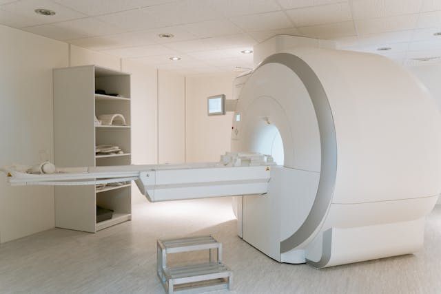
Photo by MART PRODUCTION: https://www.pexels.com/photo/photo-of-ct-scan-medical-equipment-7089013/
Sources
https://www.medicalnewstoday.com/articles/153201#uses
https://en.wikipedia.org/wiki/CT_scan
https://www.hopkinsmedicine.org/health/treatment-tests-and-therapies/computed-tomography-ct-scan
https://en.wikipedia.org/wiki/Tomography
https://www.hopkinsmedicine.org/health/treatment-tests-and-therapies/xrays
https://www.health.harvard.edu/cancer/radiation-risk-from-medical-imaging
Pingback: #43 How does an MRI work?
Pingback: #75 How does a QR code work?