
How does an electroencephalogram work? By measuring electrical activity in different parts of the brain using electrodes on the scalp.
EEGs are used to diagnose disorders with the electrical activity in the brain. These can be epilepsy, sleep disorders, anesthesia, coma, and even brain death. It is most commonly used for diagnosing epilepsy, which is caused when neurons in part of the brain start firing excessively and in synchronization. Because so many neurons in one area fire at the same time, the messages that the brain is trying to send to the rest of the body get confused and the movement of the muscles in the body can become random, causing a seizure.
The brain runs on electricity. Every thought, every movement that the brain initiates comes about because of electricity. Stimuli that the body feels, either in the ears when we hear or in the skin when we itch, are converted to electrical signals that travel up our nerves into the brain. These electrical signals are carried along the neurons in particular parts of the brain. We have about 86 billion neurons, and they all have gaps between them called synapses. When an electrical signal reaches one neuron, it can either be passed across the synapse to another neuron or stopped. This is done using chemicals. If the electrical signal is passed on, it will travel through that neuron and the process is repeated. The combinations of these signals result in everything that we feel, do, and experience. The synapses can also be affected by drugs such as painkillers, nicotine, alcohol, and caffeine.
An EEG looks at these brain signals by reading the electrical voltage of the brain in certain places. It obviously cannot read the signal from one specific neuron. It is designed to read the electrical signals from areas of the brain. Because doctors know what regular electrical signals should look like, they can tell if there are any irregularities in the electrical signals of the brain. In order to do this, the person undergoing the test will be asked to open and close their eyes or breathe in and out. They are also asked to lie quietly to gain a baseline. When various stimuli are presented, their brains will react. Doctors can compare the readings to those of a healthy patient and see what is different. By knowing which electrode read the electrical signal with the irregularity, they can work out which part of the brain is causing the problems.
During an EEG test, the electrodes are placed on the skull, usually using a conductive gel. When brain activity happens, the electrical current is transmitted through the brain, through the skull, and is picked up by the electrode. It is a tiny signal and has to be amplified, sometimes as much as 100,000 times, in order to get a reading. This is then transposed onto paper or, these days, digitally.
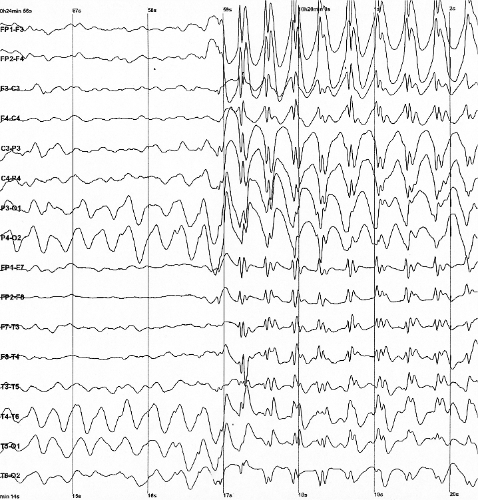
A standard EEG uses between 10 and 20 electrodes placed around the skull. The electrodes are placed over the frontal lobe, the temporal lobe, the parietal lobe, and the occipital lobe. These parts of the brain all do different things. The frontal lobe is in charge of executive function. It controls physical movement, speech control, expression of emotions, mental action, and predicting the future. The temporal lobe is used for processing sensory input of language, memories, and emotional associations. The parietal lobe is in charge of integrating information. It is responsible for hand eye coordination, amongst other things. The occipital lobe is the visual processing center of the brain. More electrodes can be used for a more detailed picture of the brain, but it becomes difficult to analyze exactly where in the brain the signals originated from. Some researchers have shown that advanced EEG technology might be able to show the presence of Alzheimer’s Disease.
Before the discovery of electricity, people had thought that the nerves carried a liquid that dripped onto nerves to stimulate them. They were unable to find the liquid but, not knowing what electricity was, they could not find any other solution either. In the early 17th century, William Gilbert used the word electricus to describe the attractive properties of charged amber. Then, in 1646, Thomas Browne coined the word electricity. Studies in electricity continued and in 1770, Luigi Galvani realized that electricity could make the body’s muscles move and work on its nerves. In 1875, Richard Caton was able to measure the electrical brain activity of rabbits and monkeys. This was followed by Adolf Beck who put sensors directly on the brains of animals to record the brain waves when they were stimulated. The first human EEG recording was made in 1924. In the 1960s, EEGs were used to monitor the brainwaves of astronauts.
So, how does an EEG work? By detecting the electrical activity in the brain and representing it in a way that a trained professional can see if there are any anomalies. If there are, it indicates which part of the brain they came from. And this is what I learned today.
Sources
https://www.epsyhealth.com/seizure-epilepsy-blog/what-happens-to-the-brain-during-a-seizure
https://en.wikipedia.org/wiki/Electroencephalography
https://www.hopkinsmedicine.org/health/treatment-tests-and-therapies/electroencephalogram-eeg
https://imotions.com/blog/what-is-eeg/
https://www.brainfacts.org/core-concepts/how-neurons-communicate
https://neuroscientificallychallenged.com/posts/history-of-neuroscience-luigi-galvani
https://en.wikipedia.org/wiki/Luigi_Galvani
https://en.wikipedia.org/wiki/Electricity
https://en.wikipedia.org/wiki/Thomas_Browne
https://en.wikipedia.org/wiki/10%E2%80%9320_system_(EEG)
https://en.wikipedia.org/wiki/William_Gilbert_(physician)